History
NUCLEAR MEDICINE AND MOLECULAR IMAGING - HISTORY
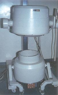
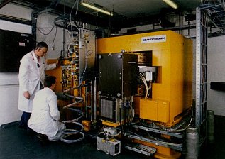
The University Medical Center Groningen and University of Groningen are pioneers in the application of radio-isotopes in medicine. This aspect of their history started in 1952 when the first in vivo and in-vitro measurements were performed. For in vivo measurements, external detectors were initially employed; later, a scanning detector system was used. Radio-immunoassays were developed. The first gamma camera was installed in 1965 and with the availability of the 99mTc generator, new in-vivo diagnostic procedures were set up. Positron Emission Tomography (PET) became possible in 1972 after installation of a large cyclotron at the Kernfysisch Versneller Insituut (KVI), a university institution. Pioneering PET studies were performed in a close cooperation between Nuclear Medicine, the KVI and the University Laboratory of Organic Chemistry. This led to financing by the Dutch government of a fully equipped PET-Center on hospital grounds in 1988. Nuclear Medicine (headed by Dr.D.A.Piers) and PET-Center (headed by Prof.W.Vaalburg) developed via separate lines during the 1990s. Because of the clinical potential of nuclear imaging in diagnostics and radionuclide therapy, Nuclear Medicine and PET-Center were re-united in 2005. The Department of Nuclear Medicine and Molecular Imaging was formed as a novel discipline group of the UMCG, headed by Prof.Dr. R.A.Dierckx