Medical Visualisation
3D reconstruction
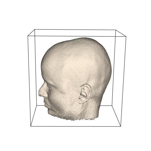
The term Medical Visualisation applies in particular to the 3D reconstruction of stacks of CT- of MRI-images. The first picture was reconstructed from a stack of 162 256*256 MRI-images using the isosurface technique of the Visualisation Toolkit (VTK).

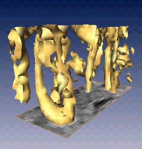
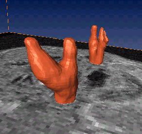
By cutting large parts away from the head, we can view in the interior of the head to observe the veins in the lower part, picture below. In the third picture we see the veins segmentated by Amira.


A journey though the throat
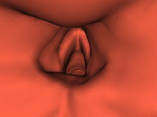
Using the same isosurface technique of VTK, a stack of 161 CT-images of the human throat was visualised. By forming images from points along a line through the throat, we constructed a movie that suggests a fly through the throat. Click an image to activate the movie!
3D -reconstructions can be used for diagnosis and surgical traning.
See also PR-leaflet.
Some (Dutch) links
- Aanwending van virtuele realiteit in de operatiezaal: een klinische toepassing uit de cardiologie (KU Leuven)
- Home page of Dirk Bartz, Tuebingen: virtual endoscopy, volume rendering, parallel computing, large model visualisation, ...

| Laatst gewijzigd: | 04 oktober 2024 12:41 |
